Sara Cermeno Fernandez
Resident in Small Animal Medicine
LV MRCVS
Hypercalcaemia is a common cause of polydipsia/polyuria, hyperoxia and lethargy in dogs. Treatment and prognosis entirely depend on the underlying cause and therefore a logical work-up is important.
Calcium presents within the circulation in three forms – ionised calcium (50%), which is the active form; protein-bound calcium (40%) and the chelated form complexed with lactate, citrate and bicarbonate (10% of total calcium).
Ionised calcium needs to be measured in order to identify the true status of calcium within the body. This is the physiologically active form which is normally kept within a tight reference range. Ongoing hypercalcaemia can cause kidney damage, and also increases the risk of urolithiasis. Hypercalcaemia needs to be investigated further in order to start appropriate treatment.

Clinical signs associated with hypercalcaemia include:
- Polyuria and polydipsia
- Lethargy, weakness, poor appetite and weight loss
- Uncommon clinical signs include vomiting, diarrhoea, constipation and arrhythmias. Seizures can occur in severe cases
- The most common clinical signs in cats are anorexia and lethargy, while polydipsia and polyuria are less common
Differential Diagnosis
The most common causes of hypercalcaemia in dogs are malignancy (lymphoma and anal sac adenocarcinoma are the two most common causes), hypoadrenocorticism, primary hyperparathyroidism and chronic kidney disease.
However, malignancy and kidney disease are common causes of hypercalcaemia in cats. Furthermore, in cats, squamous cell carcinoma was associated with hypercalcaemia as frequently as was lymphoma. Idiopathic hypercalcaemia is an important differential in cats, while this is not identified in dogs.
Differential diagnoses include:
- Hyperparathyroidism
- Hypoadrenocorticism/Addisons disease
- Chronic kidney disease
- Hypervitaminosis D (Vitamin D toxicosis): Cholecalciferol rodenticide toxicosis, plant intoxication (e.g.: blooming jasmine), excessive dietary supplementation, ingestion of human medication containing calcitriol (e.g.: psoriasis cream) and over-supplementation for hypoparathyroidism are causes reported for hypercalcaemia
- Idiopathic (Cats)
- Osteolytic disease
- Neoplasia - Lymphoma, apocrine gland adenocarcinoma of the anal sac and multiple myeloma are common paraneoplastic causes of hypercalcaemia. Other tumours such as squamous cell carcinoma, lung carcinoma, malignant melanoma, osteosarcoma, metastatic bone tumours and mammary carcinoma are less common causes of hypercalcaemia
- Granulomatous disease
- Spurious - lab error or hyperproteinaemia are causes for increased calcium levels
- Young age - total hypercalcaemia can be normal in young dogs, however ionised calcium levels should be within normal limits
Diagnostic Approach
History should be obtained including duration of clinical signs, any changes to drinking, energy levels or behaviour and potential access to toxins, medication or diet for possible hypervitaminosis D. A thorough physical examination is very important to carefully assess peripheral lymph nodes and a rectal examination should also be performed.
Investigations:
- CBC and biochemistry – in order to check albumin levels, kidney parameters, electrolytes and phosphate
- Urinalysis including urine culture
- Basal cortisol measurement +/- ACTH stimulation test if cortisol <55nmol, to rule out hypoadrenocorticism
- PTH and PTHrp level analysis is necessary to confirm diagnosis of suspected hyperparathyroidism
- Imaging including three view thoracic radiographs, abdominal ultrasound or CT scans are usually needed to screen for common causes of hypercalcaemia. Fine needle aspirates of any tissue abnormalities are advised for cytology. Biopsies should be considered in some cases
- Measurement of vitamin D is recommended in patients with history consistent with hypervitaminosis D
- ECG and echocardiography should be evaluated when arrhythmia is present
Medical Treatment
Acute therapy
This is often guided by the severity of clinical signs rather than using a specific cut-off value. Ultimately removal or effective treatment of the underlying cause is the definitive treatment for hypercalcaemia, but this is not always immediately possible.
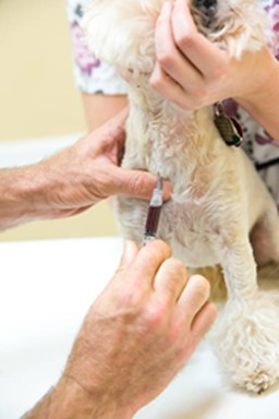
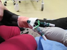
The goals for initial medical management of hypercalcaemia are to enhance renal excretion of calcium and prevent calcium re-absorption. Fluid therapy with 0.9% sodium chloride can restore fluid volume and correct dehydration, as well as induce diuresis. Electrolytes should be monitored as potassium supplementation can be necessary in some cases. Furosemide can be used to increase diuresis in hydrated patients if clinical signs are present.
Chronic treatment
If the underlying cause can’t be treated, e.g.: unresectable neoplasia or idiopathic hypercalcaemia in cats, then additional treatment can be trialled. Glucocorticoids are effective in reducing calcium levels in the case of malignancies, hypervitaminosis D and granulomatous disease, although it is recommended to withhold corticoid therapy until a definitive diagnosis is made. Steroids will decrease intestinal calcium absorption, decrease renal tubular calcium reabsorption and decrease skeletal mobilisation of calcium. Bisphosphonate can be used intravenously or orally, but the latter is poorly absorbed intestinally and therefore often less effective
Dietary Management
Dietary management is generally used for chronic cases of idiopathic hypercalcaemia. A high fibre diet, e.g. Royal Canin Fibre Response™ or Hills W/D™ reduces calcium reabsorption from the gastro intestinal tract.